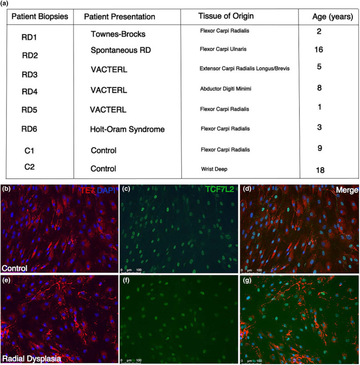
FIGURE 1.

Connective tissue fibroblast characterisation. Patient biopsy samples are listed alongside the patient presentation and the tissue of origin that the biopsies are obtained (a). Control (b–d) and radial dysplasia (e–g) patient connective tissue cells are characterised using targeted fibroblast (TE7) and muscle connective tissue (TCF7‐like 2) antibody staining. Most of the cultured cells from the patient biopsies highly express TE7 and TCF7‐like 2, suggesting the population of cells are connective tissue fibroblasts (b–g).
