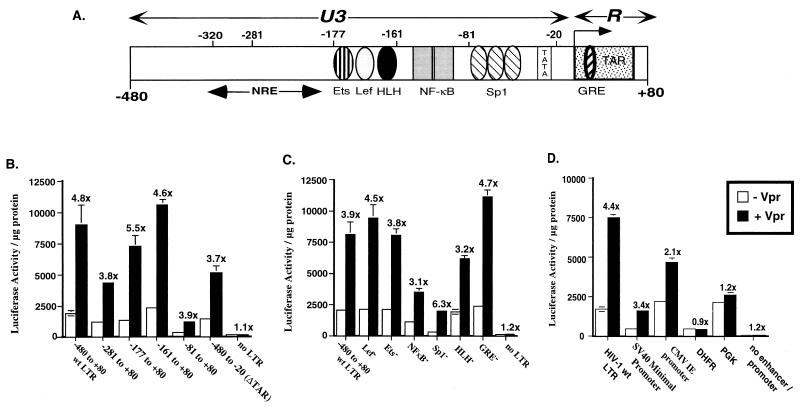
FIG. 5.
Delineation of HIV-1 LTR sequence requirements for Vpr-mediated stimulation of the HIV-1 LTR. (A) Schematic representation of the HIV-1 LTR. Positions of the transcription factor binding sites that were assayed in the transfection experiments, the TATA box, and the TAR element are marked. NRE, negative regulatory element. Jurkat cells were cotransfected with 0.625 μg of expression plasmid pCMV-Vpr and 0.625 μg of either sequentially deleted LTRs (B); LTRs with mutations in the individual transcription factor binding sites including Lef, Ets, NF-κB, Sp1, helix-loop-helix (HLH) protein binding motif, and GRE (C); or viral (HIV-1 LTR, SV40 minimal promoter, and CMV IE promoter) and cellular (human DHFR and mouse PGK-1) promoters driving luciferase expression (D). Names of the plasmids in panel B identify the portion of the HIV-1 LTR included in each construct, and the nucleotide positions are denoted with respect to the +1 transcription start site. In all transfections, the empty CMV expression plasmid was cotransfected with each reporter plasmid as a control. Luciferase assays were performed on cell lysates harvested 48 h posttransfection, and results were expressed as luciferase activity in relative light units per microgram of protein (y axis). Fold increase in luciferase activity obtained in the presence of Vpr over the luciferase activity obtained in the absence of Vpr is indicated above the histograms, and error bars represent standard errors of mean. Reported values are means of transfections performed in triplicate. The experiment has been repeated at least three times with similar results. wt, wild type.

