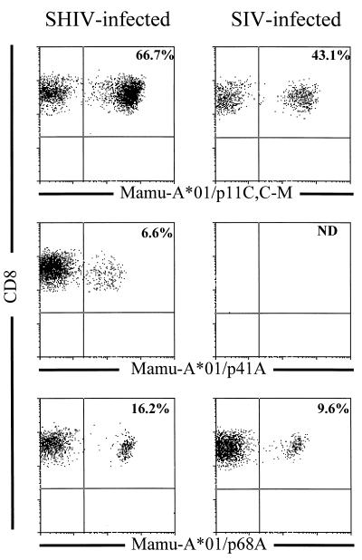
FIG. 4.
Tetramer binding to peptide-stimulated PBMC from SHIV- and SIVmac-infected, Mamu-A*01+ rhesus monkeys. PBMC from SHIV-89.6-infected monkey 287 and SIVmac-infected monkey 4DK were stimulated in vitro with 1.0 μg of the indicated optimal peptide (p11C, C-M [CTPYDINQM], p68A [STPPLVRLV], or p41A [YAPPISGQI]) per ml for 10 days in rIL-2-containing medium. The PE-coupled tetrameric Mamu-A*01/peptide/β2m complexes were used in combination with anti-CD8α-FITC, anti-CD8αβ-ECD, and anti-CD3-APC to stain 2 × 10⁵ lymphocytes isolated by Ficoll-Hypaque density gradient centrifugation following this in vitro peptide stimulation. The percentage of CD8+ T lymphocytes staining positively with the Mamu-A*01/peptide/β2m complex is indicated in the upper right quadrant.

