
Fig. 4 |. Revealing in situ clonal tracing by mtDNA genetic variation.
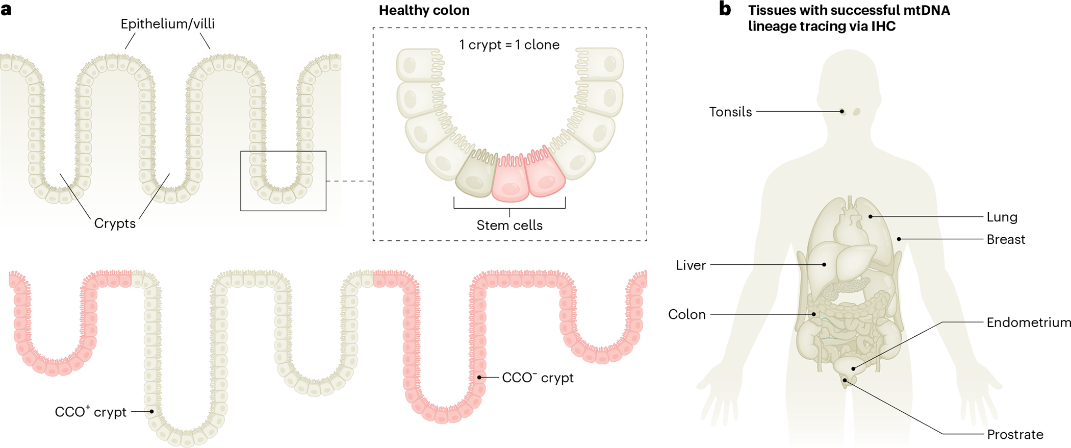
a, Schematic illustrating how stem cells in colonic crypts may acquire somatic mtDNA mutations in cytochrome c oxidase (CCO), leading to the loss of biochemical activity (pink) compared to wild-type cells (beige), as measured by immunohistochemistry (IHC). As mtDNA mutations may be propagated across cell divisions, all stem-cell-derived daughter cells are ‘visually’ marked as they all exhibit a lack of CCO activity, thereby facilitating the identification of clonally related cells. b, Overview of human organ systems surveyed with classical immunohistochemical or immunofluorescence approaches to identify clonal patches of cells on the basis of the loss of protein activity or expression of mtDNA-encoded genes.