Figure 2.
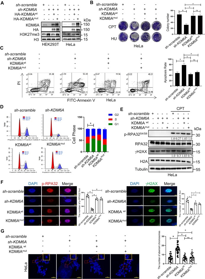
KDM6A regulates the genomic stability independently on its demethylation activity. (A) Western blot with the indicated antibodies in HEK293T and HeLa cells knocked down endogenous KDM6A and followed by transfection with the plasmid of KDM6Awt or catalytic-dead KDM6Amut respectively. (B) Cellular viability for genotoxin CPT or HU treated HeLa cells which is used in panel A. (C) Apoptosis determined by flow cytometry for HeLa cells used in panel A in the presence of CPT. (D) Cell cycle profile for HeLa cells used in panel A. (E) Western blot for phospho-RPA32 (S4/S8) and γH2AX in HeLa cells used in panel A. Phospho-RPA32 (S4/S8) and γH2AX expression were normalized to RPA32 and γH2AX expression respectively and the quantification number was labeled below the protein bands. (F) Image of phospho-RPA32 (S4/S8) and γH2AX foci was captured in HeLa cells used in panel A by fluorescence microscopy. Representative images with scale bars representing 5 μm and quantifications are shown. (G) Abnormal chromosome for the indicated HeLa cells. Representative images and quantifications are shown. Scale bars, 5 μm. All experiments were independently repeated three times, *P < 0.05. P values were determined using a paired Student's t test.

