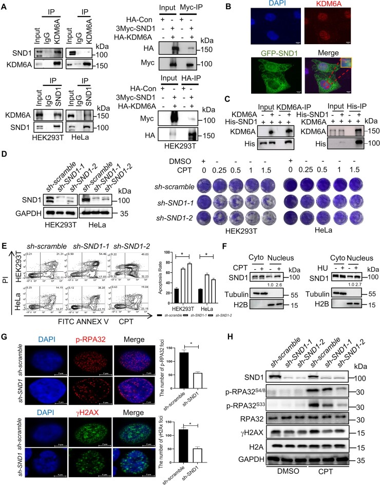
Figure 4.
SND1 is an interactor protein of KDM6A and involved in the genomic stability. (A) The interaction between KDM6A and SND1 in vivo. KDM6A and SND1 was immunoprecipitated using specific antibodies against KDM6A and SND1 respectively or specific tag antibodies, followed by western blot with indicated antibodies. (B) The co-localization between KDM6A and SND1 in HeLa cells. Representative images with scale bars (5 μm) and quantifications are shown. (C) Detection of KDM6A–SND1 binding in vitro using purified recombinant proteins. In vitro pull-down assay was performed using purified KDM6A and His-SND1 recombinant proteins followed by detection with western blot. (D) The validation of SND1 knockdown by western blot (Left panel) and cellular viability against CPT in HEK293T and HeLa cells (Right panel). (E) Apoptosis for indicated cells with SND1 knockdown in the presence of CPT. (F) Western blot for the chromatin associated KDM6A and SND1. Chromatin-associated proteins were prepared from the nucleus of HEK293T cells treated with either CPT or HU and were subjected to western blot. The number represents the quantification of KDM6A against to H2B. (G) Image of phospho-RPA32 (S4/S8) and γH2AX foci in HeLa cells was captured by fluorescence microscopy. Representative images with scale bars (5 μm) and quantifications are shown (left). (H) Western blot for phospho-RPA32 (S4/S8 and S33) and γH2AX in HeLa cells. All experiments were independently repeated three times (right panel). *P < 0.05. P values were determined using a paired Student's t test.

