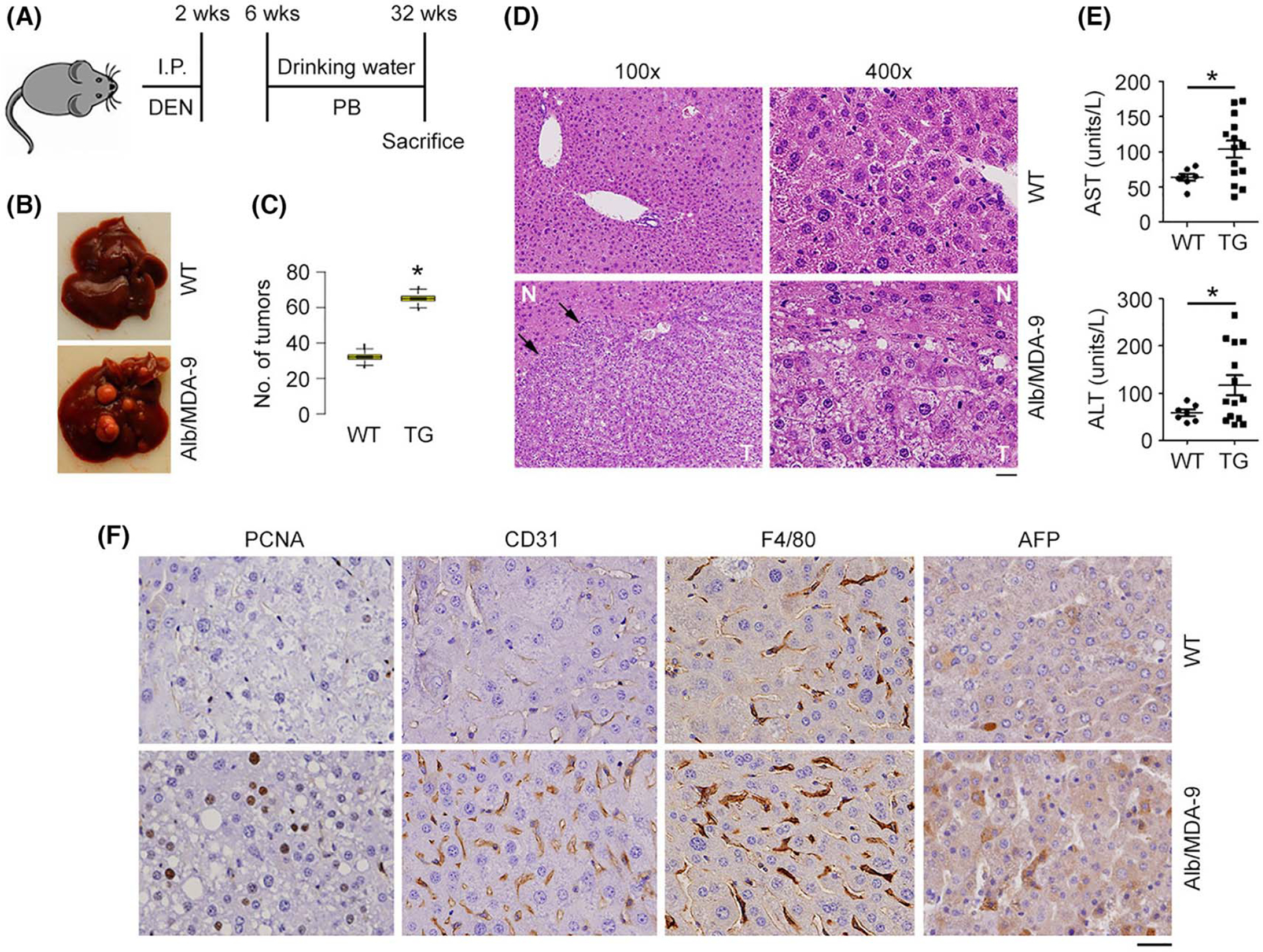
FIGURE 2.

HCC is augmented in Alb/MDA-9 mouse. (A) Schematic of experimental protocol. WT and Alb/MDA-9 littermates were injected with the hepatocarcinogen DEN (10 μg/g bw) at 2 weeks of age, and starting at 6 weeks of age, they were given PB (0.05%) in drinking water, which serves as a mitogen. The mice were sacrificed at 32 weeks. (B) Representative photograph of DEN/PB-treated WT and Alb/MDA-9 livers at 32 weeks of age. (C) Number of tumors in WT and Alb/MDA-9 (TG) livers. Data represent mean ± SEM. *p < 0.01. (D) H&E staining of DEN/PB-treated WT and Alb/MDA-9 liver sections at 32 weeks of age. Arrows indicate tumor margin. Scale bar: 20 μm. (E) AST and ALT levels in the sera of DEN/PB-treated WT and Alb/MDA-9 (TG) mice at 32 weeks of age. Data represent mean ± SEM. *p < 0.01. (F) Representative IHC staining of the indicated markers in WT and Alb/MDA-9 liver tumors. Magnification: 400×. Scale bar: 20 μm. AFP, alpha-feto protein; Alb, albumin; ALT, alanine aminotransferase; AST, aspartate aminotransferase; DEN, N-nitrosodiethylamine; H&E, hematoxylin and eosin; HCC, hepatocellular carcinoma; IHC, immunohistochemistry; I.P., intraperitoneal; MDA-9, Melanoma differentiation associated gene-9; PB, phenobarbital; TG, transgenic; WT, wild-type.
