
Figure 1.
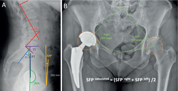
(A) Lateral standing spinopelvic radiographs and (B) AP pelvic radiograph illustrate the measurements performed; LL = Cobb angle between a line drawn along the superior endplate of L1 and another line drawn along the superior endplate of S1; SS = angle between a line drawn along the superior endplate of S1 and the horizontal axis; PI = angle between the line from the center of the cup to the middle of the superior endplate of S1 and the line perpendicular to the superior endplate of S1 from its midpoint; PT = angle formed between the line from the center of the cup to the middle of the superior endplate of S1 and the vertical axis; PFA = angle between the line from the center of the cup to the middle of the superior endplate of S1 and the femoral axis; APP = angle between a line connecting both anterior superior iliac spines with the pubic symphysis and the vertical axis; SFP = angle between a line from the midpoint of the S1 superior endplate (found by determining the midpoint of a line between the lateral bodies of the L5 to S1 facet joints), centroid of the acetabulum, or center of the cup, and upper midpoint of the pubic symphysis.