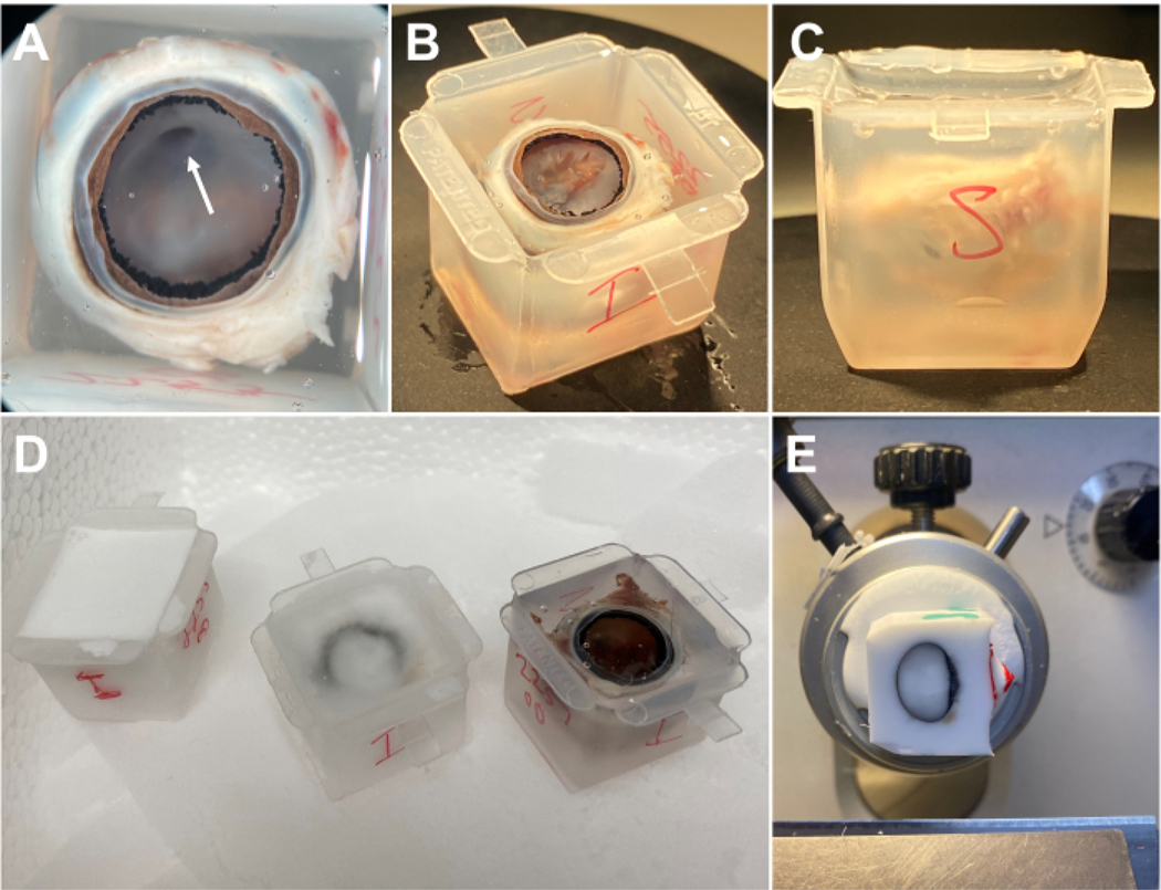
Figure 2: Embedding of rabbit eye.
(A) After overnight incubation in OCT, the hyaloid membrane should appear lifted. The hyaloid canal (arrowhead) may be visible and can be used for orientation purposes. (B) For better orientation, the hyaloid membrane may be dissected to reveal the optic nerve head underneath. (C) The eye should not touch any part of the cryomold and should be sufficiently submerged without significant bubble formation. (D) Cryomolds should be placed on dry ice. The appearance at various stages of freezing is shown. (E) After freezing, the block may be sectioned in the cryostat. This block is oriented with the pupil facing rightward. OCT is visible within the eye and in close apposition to the inner and outer surface of the eye. Abbreviation: OCT = optimal cutting temperature.

