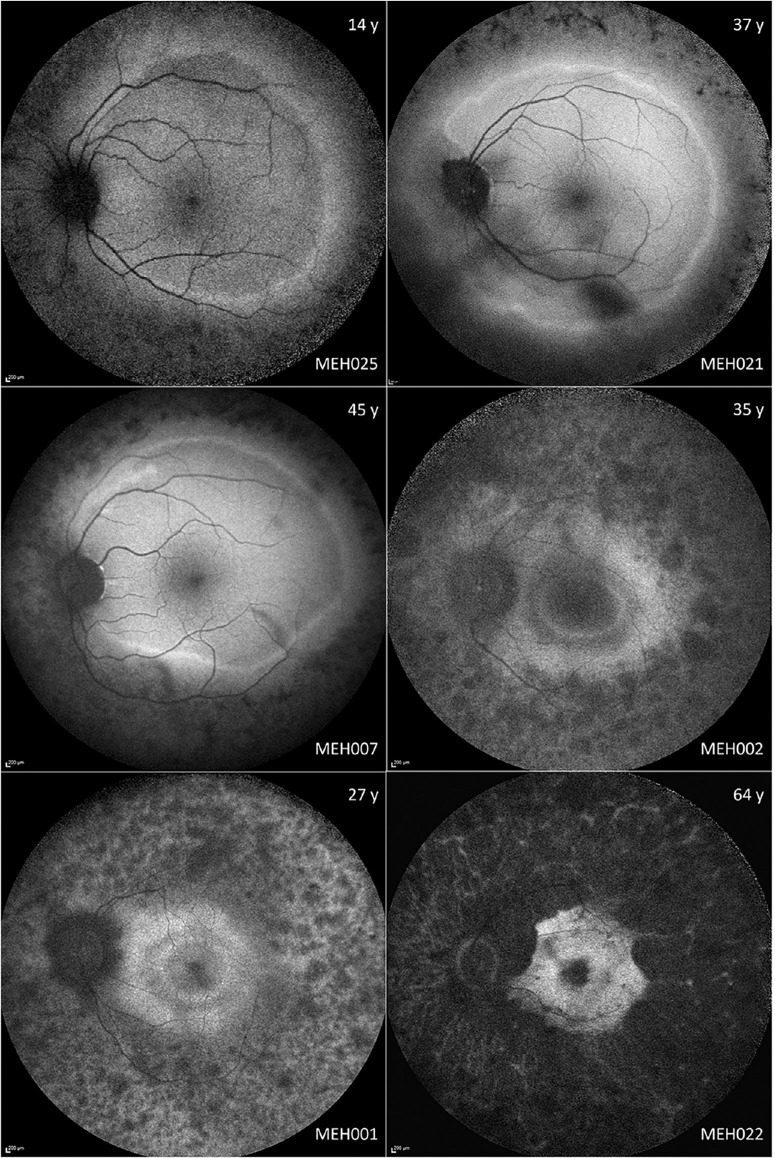
Figure 3.
Montage of fundus autofluorescence (FAF) imaging in different disease stages. On the top and bottom right corner are the age (years) and the subject ID, respectively. The most frequent early imaging findings are hyperautofluorescent rings, which are usually located beyond the vascular arcades. With disease progression, the rings tend to gradually constrict. MEH002 and MEH001 have double hyperautofluorescent rings separated by a patch of normal autofluorescence.

