
Figure 6. Activation mechanism of CLC-ec1.
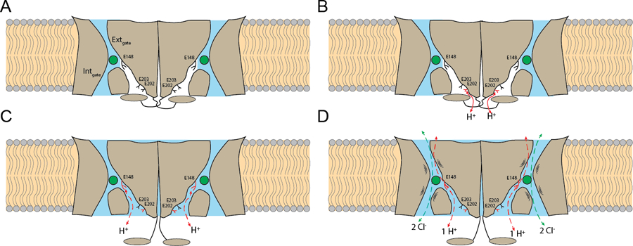
(A) The transporter is inactive. The H+ pathways are dehydrated (in white) and occluded by the cytosolic region (in gray), E148, E202 and E203 are deprotonated (in black), and the Cl− pathways (in blue) are constricted at the internal and external gates. (B) E202 is partially exposed to the intracellular solution and becomes protonated (in red). (C) The cytosolic regions disengage from the transmembrane regions and open the H+ pathways, that become hydrated (in blue) allowing formation of short water wires between E148 and the intracellular solution (red dashed arrows). (D) The Cl− pathways become dynamic, with openings of the internal or external gates to allow Cl−:H+ exchange to occur.