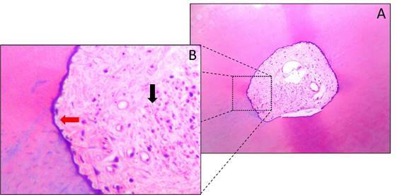
Figure 2. Representative photomicrographs of the cross sections of the apical region of mandibular premolars from the control group (without instrumentation or irrigation). (A) 100x magnification. (B) 400x magnification; note the presence of organic and inorganic tissue (black arrow) and intact walls (red arrow).

