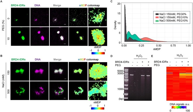
Fig. 2.
Phase-separated BRD4-IDRs minimize the DNA damage in vitro. (A) BRD4-IDRs were diluted in the buffer to a final concentration of 5 μM in the presence of DNA (5 ng/μl) and indicated conditions. Colocalization in ROI was visualized using nMDP colormap. (B) BRD4-IDRs and DNA were diluted in the buffer to a final concentration of 5 μM and indicated conditions in the presence of 10% PEG. (C) The extent of colocalization was quantified using nMDP value and visualized by density plot. (D) Result of DNA damage represented in the presence of BRD4-IDRs with/without 10% PEG. (E) Heatmap visualization of the extent of DNA amounts. Maximum signal of DNA (non-damaged DNA) was prepared as 100%.

