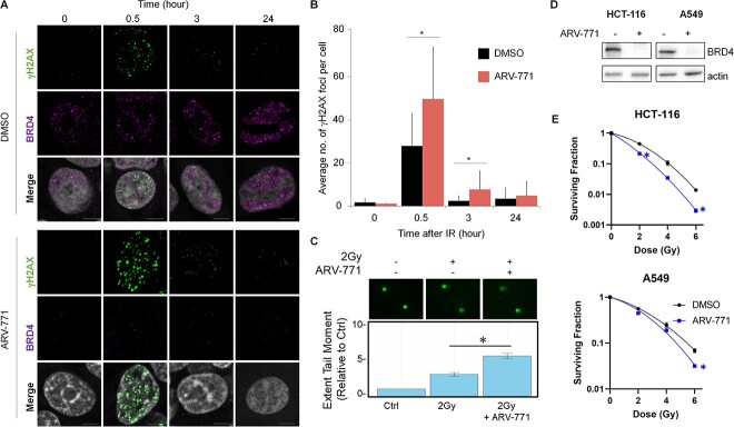
Fig. 4.
Pharmacological degradation of BRD4 works as radiation sensitizer. (A) Representative images of BRD4 and IR-induced γH2AX in the cells with either 2 Gy IR alone or a combination of IR and BRD4 degradation. (B) Quantification of γH2AX foci (30 cells) based on a single experiment. Similar results were obtained in two independent experiments. Data show mean ± SD. (C) Fluorescence microscopy images of neutral comet assay in cells upon 2 Gy pretreated for 12 h with DMSO or ARV-771 (n = 3). The bar graph shows mean ± SEM. Significance was assessed using a Student’s t-test (*P < 0.05). (D) Western blotting analysis of BRD4 upon ARV-771 treatment in HCT116 and A549 cell lines. (E) Survival fraction of HCT116 and A549 cell lines upon IR alone and IR combined with ARV-771.

