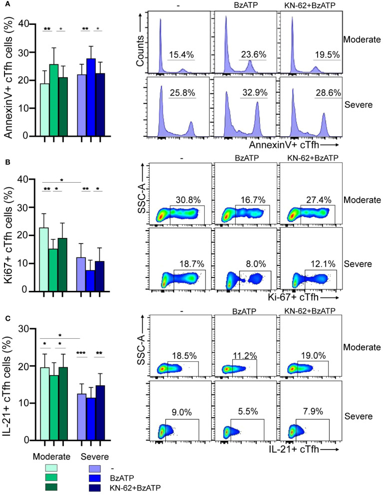
Figure 4.
Modulation of viability and function of cTfh cells via P2X7R stimulation during RSV infection. (A) Left: PBMCs (1x10⁶/mL) from RSV moderate (n=9) and severe children (n=8) were incubated with BzATP (300 µM), BzATP plus KN-62 (1µM) or nontreated for 24 hours. Percentage of apoptosis of cTfh cells was analyzed by flow cytometry. Right: Representative histograms showing Annexin V+ cTfh cells in a donor of each cohort are depicted. (B) Left: PBMCs (1x10⁶/mL) from RSV moderate (n=10) and severe children (n=8) were stimulated with anti-CD2/CD3/CD28 coated beads (0.75 μg/mL) and treated or not with BzATP (100 μM) and/or KN-62 (1µM) and cells were culture for 3 days. Frequency of cTfh Ki-67+ cells was evaluated by flow cytometry. Right: Representative dot plots showing Ki-67+ cTfh cells in a donor of each cohort are shown. (C) Left: PBMCs from RSV moderate (n=9) and severe children (n=10) were treated or not with BzATP (100 μM) and/or KN-62 (1µM) for 24 hours. Afterward, were re-stimulated with PMA and Ionomycin in the presence of monensin for 5 hours. Percentage of cTfh IL21+ cells were analyzed by flow cytometry. Right: Representative dot plots showing IL-21+ cTfh cells in a donor of each cohort are depicted. Mean ± SEM of n donors are shown in A (left), B (left) and C (left). P values were determined by Wilcoxon, Friedman test followed by Dunn’s multiple comparison test and Mann-Whitney U test. *p<0.05, ** p<0.01, *** p<0.001. Moderate (green squares), severe (blue squares).

