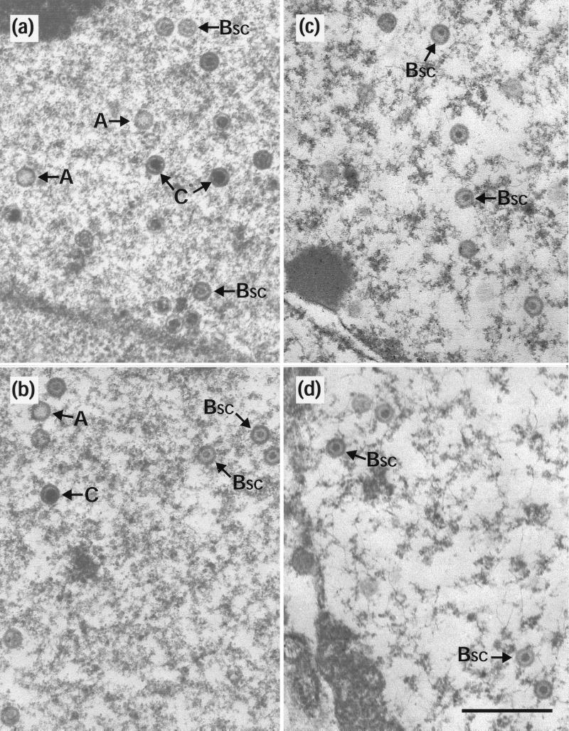
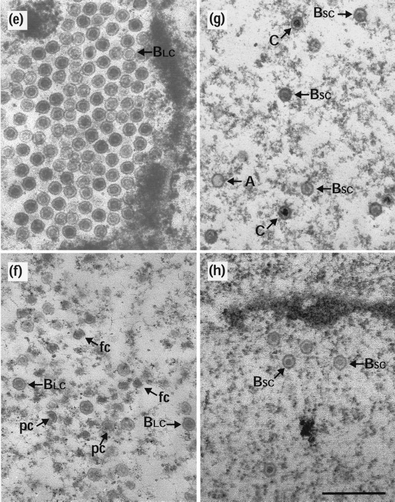
FIG. 2.
Capsid stability at 0°C. Replica 35-mm-diameter plates of BHK C13 cells were infected with 5 PFU of HSV-1 wt virus (a and b), ts1203 (c and d), or ts1201 (e to h) per cell. After incubation at 39°C for 10 h, the plates were either harvested immediately for electron microscopy (a, c, and e) or overlaid with fresh prewarmed or precooled medium containing 200 μg of cycloheximide per ml and incubated further at the desired temperature. For both wt (b) and ts1203 (d), cells were overlaid with medium that had been precooled to 0°C and were then incubated on ice for a further 4 h. For ts1201, one plate was treated the same as for wt and ts1203 by being incubated at 0°C for 4 h (f), while two further plates were incubated at 31°C for 4 h. One of these was then harvested immediately for electron microscopy (g), and the other was overlaid with medium at 0°C and incubated for a further 10 h on ice (h). A, A capsids; C, C capsids; pc, partial capsids; fc, free cores. Scale bar = 500 nm.


