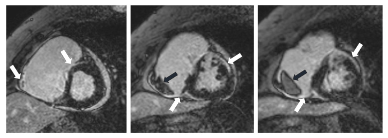
Fig. 2.
CMR images of a 70-year-old female patient with CS. The delayed contrast enhancement images in short axis planes show biventricular late gadolinium enhancement (LGE) corresponding to fibrotic involvement (white arrows) and right ventricular thrombus formation (black arrows). Subepicardial LGE is present in the anterior septum, LV inferior wall, subepicardial-midmyocardial LGE is seen in the LV anterior wall and LGE is present in the RV myocardium in the vicinity of thrombus. CMR, cardiac magnetic resonance; CS, cardiac sarcoidosis; LV, left ventricle; RV, right ventricle.

