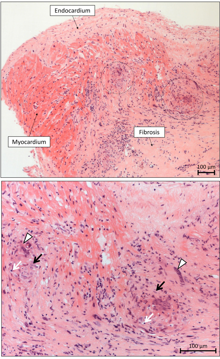
Fig. 4.
Microscopic appearance of sarcoidosis in the endomyocardial biopsy specimen. Top: The non-necrotizing granulomatous inflammation of the myocardium is sharply demarcated, and it is typically surrounded by diffuse fibrosis. The granulomas (- - -) are scattered and typically well circumscribed in sarcoidosis. Diffuse necrosis of cardiomyocytes is absent (H&E staining, 10() objective magnification). Bottom: The cellular components of sarcoid granulomas include multinucleated giant cells (), epithelioid macrophages (black arrows) and lymphocytes (white arrows) (H&E staining, 20() objective magnification).

