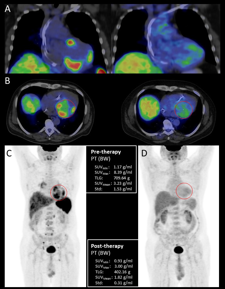
Fig. 5.

Pre- and posttreatment FDG-PET/CT examination of a 44-year-old male patient with histologically (EMB) proven sarcoidosis. Coronal fused pretreatment and posttreatment (A) and axial fused (B) PET/CT images with volume of interest (VOI) and maximum intensity projection (MIP) PET images before (C) and after immunosuppressive therapy (D) with quantitative parameters. Pretreatment scans show increased multifocal FDG uptake in the left and right ventricular myocardium as cardiac involvement, the presence of high focal supra- and infradiaphragmatic lymph node uptake is indicative of extracardiac sarcoidosis. Posttreatment scans do not show pathological FDG uptake confirming complete treatment response. EMB, endomyocardial biopsy; BW, body weight; FDG-PET/CT, 18F-fluorodeoxyglucose positron emission tomography/computed tomography; PT, positron emission tomography; SUV, standardized uptake values; TLG, total glycolytic activity.
