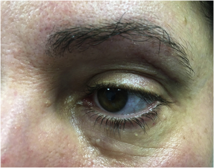
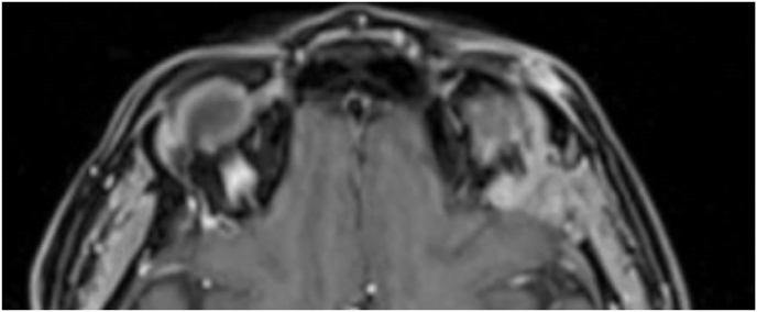
Fig. 2.
Axial (a) and coronal (b) T1-weighted gadolinium contrast-enhanced MRI of the orbit showing a hyperintense left extraconal meningioma of the lateral wall involving the orbital apex; (c) Endoscopic intraoperative photograph obtained during tumor excision. OR: orbital roof; M: meningioma; LW: lateral orbital wall; (d) Patient photograph obtained 6 months after surgery and showing a good cosmetic result in the affected eye; (e) Post-op T1-weighted gadolinium contrast.enhanced MRI of the orbit.


