Fig. 3.
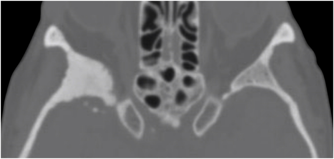
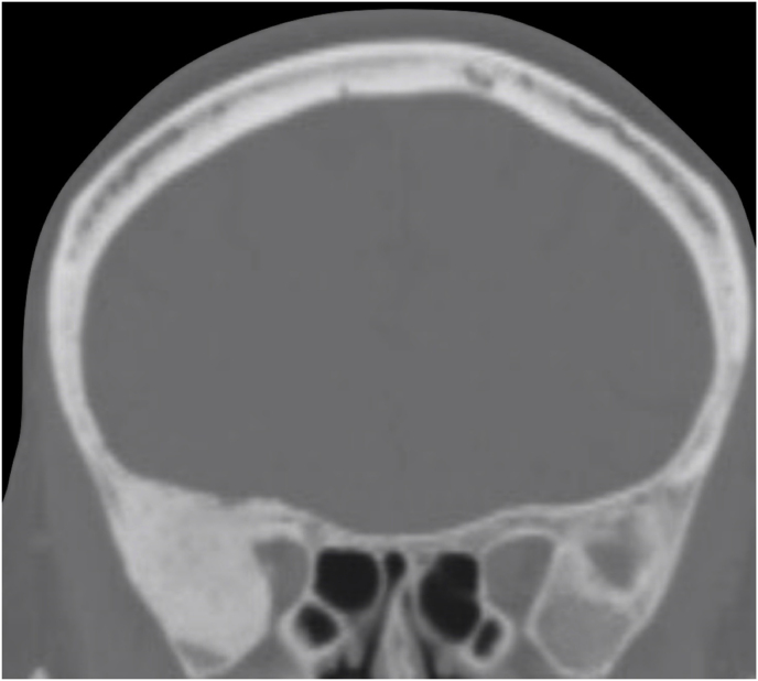
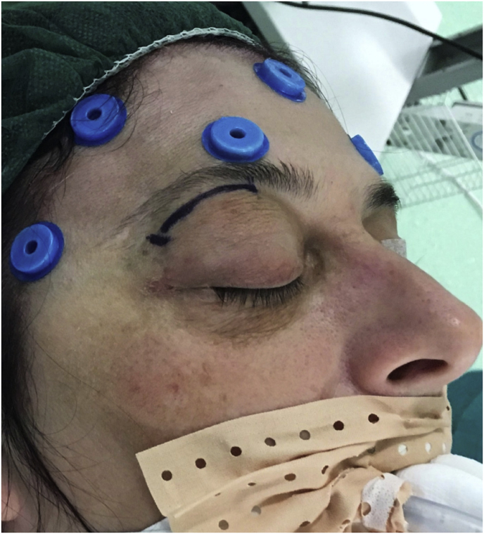
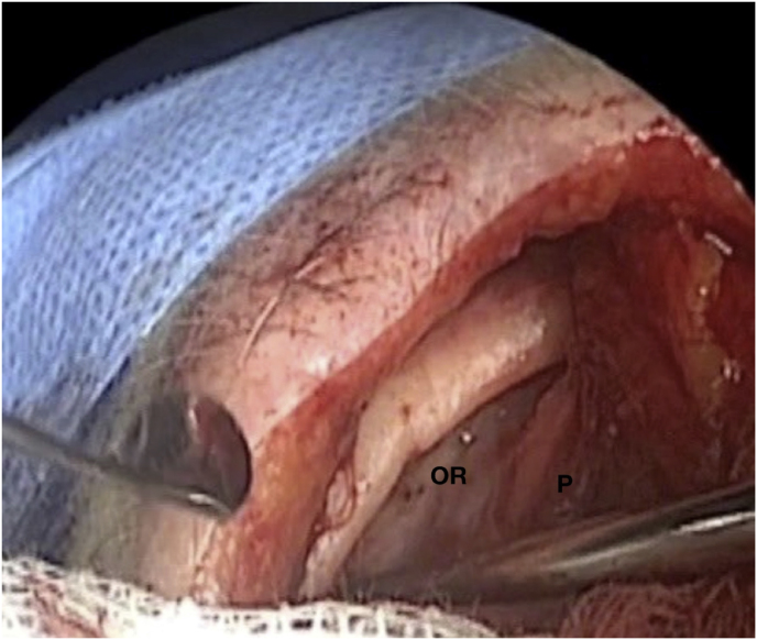
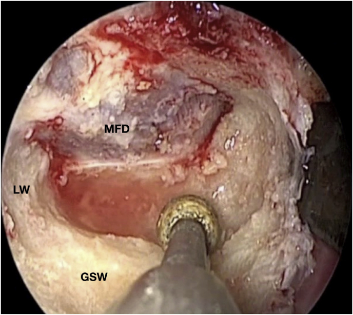
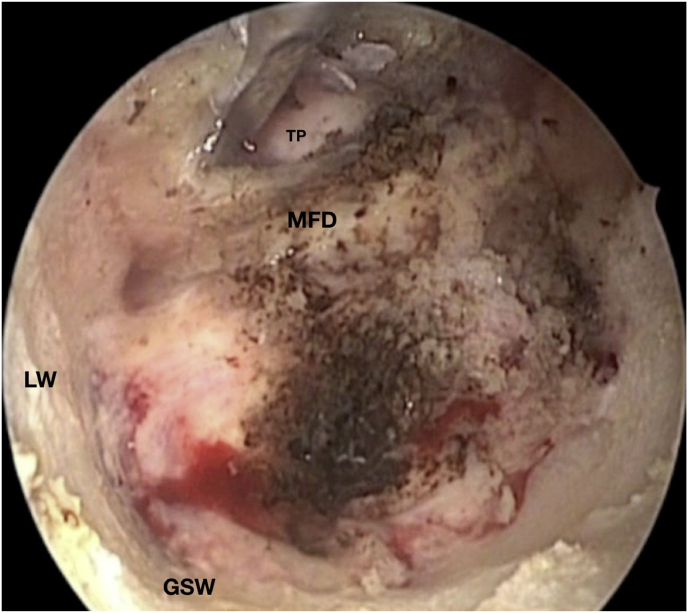
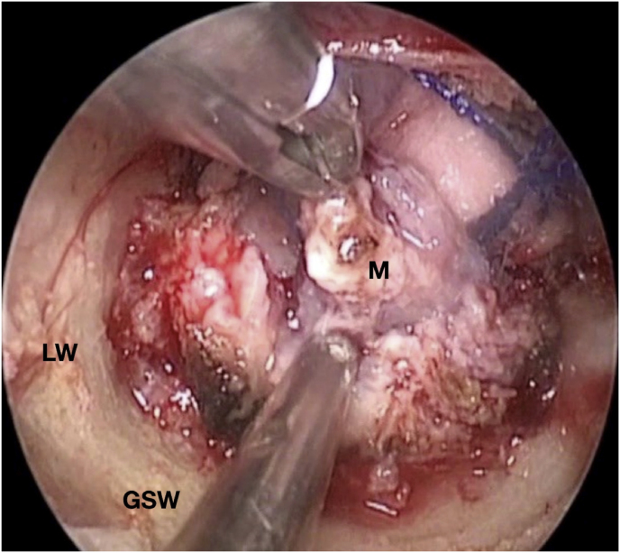
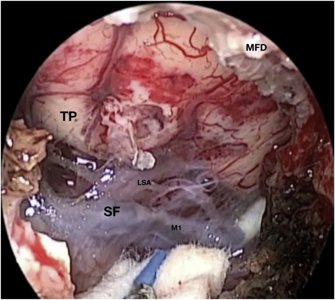
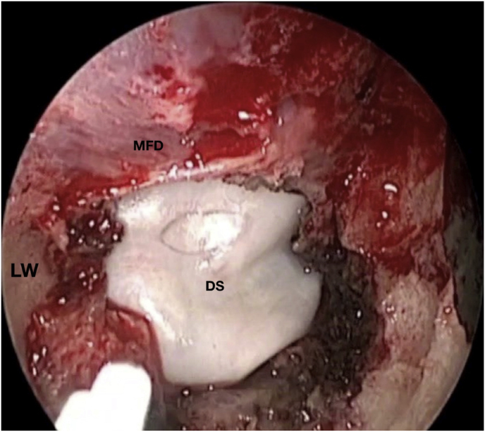
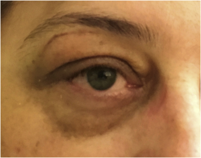
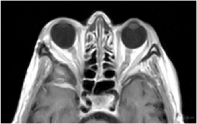
Axial (a) and coronal (b) T1-weighted gadolinium contrast-enhanced MRI of the right spheno-orbital region showing a spheno-orbital meningioma with a clear dural tail involving the orbital apex; Axial (c) and coronal (d) non-enhanced bone-window CT showing massive hyperostosis of the lateral wall of the right orbit and greater sphenoid wing; (e) Intraoperative image showing the patient placed in a supine position with the head fixed by a three-point head clamp; (f) Intraoperative image showing the subperiosteal dissection of the periorbita after skin incision; (g) Extensive drilling of the sphenoid ridge allowed exposure of the middle fossa dura; (h) the opening of the spheno-orbital dura was made just above the orbital apex, and the meningioma was fully exposed, debulked and dissected (i); (j) Intraoperative photograph at the end of surgery showing the sphenoidal compartment of the Sylvian fissure, the Sylvian cistern, the M1 segment of middle cerebral artery, along the origin of the lateral lenticulostriate arteries; (k) onlay reconstruction of the spheno-orbital dural defect with collagen dura substitute and fibrin glue; (l) image of the patient obtained 6 months after surgery revealing a good cosmetic outcome; (m) Post-op T1-weighted gadolinium contrast.enhanced MRI of the spheno-orbital region
OR: orbital roof; P: periorbita; MFD: middle cranial fossa dura; LW: lateral orbital wall; GSW: greater sphenoid wing; TP: Temporal pole; M: meningioma; SF: Sylvian fissure; M1: M1 segment of the middle cerebral artery; LSA: lateral lenticulostriate arteries; DS: dural substitute.


