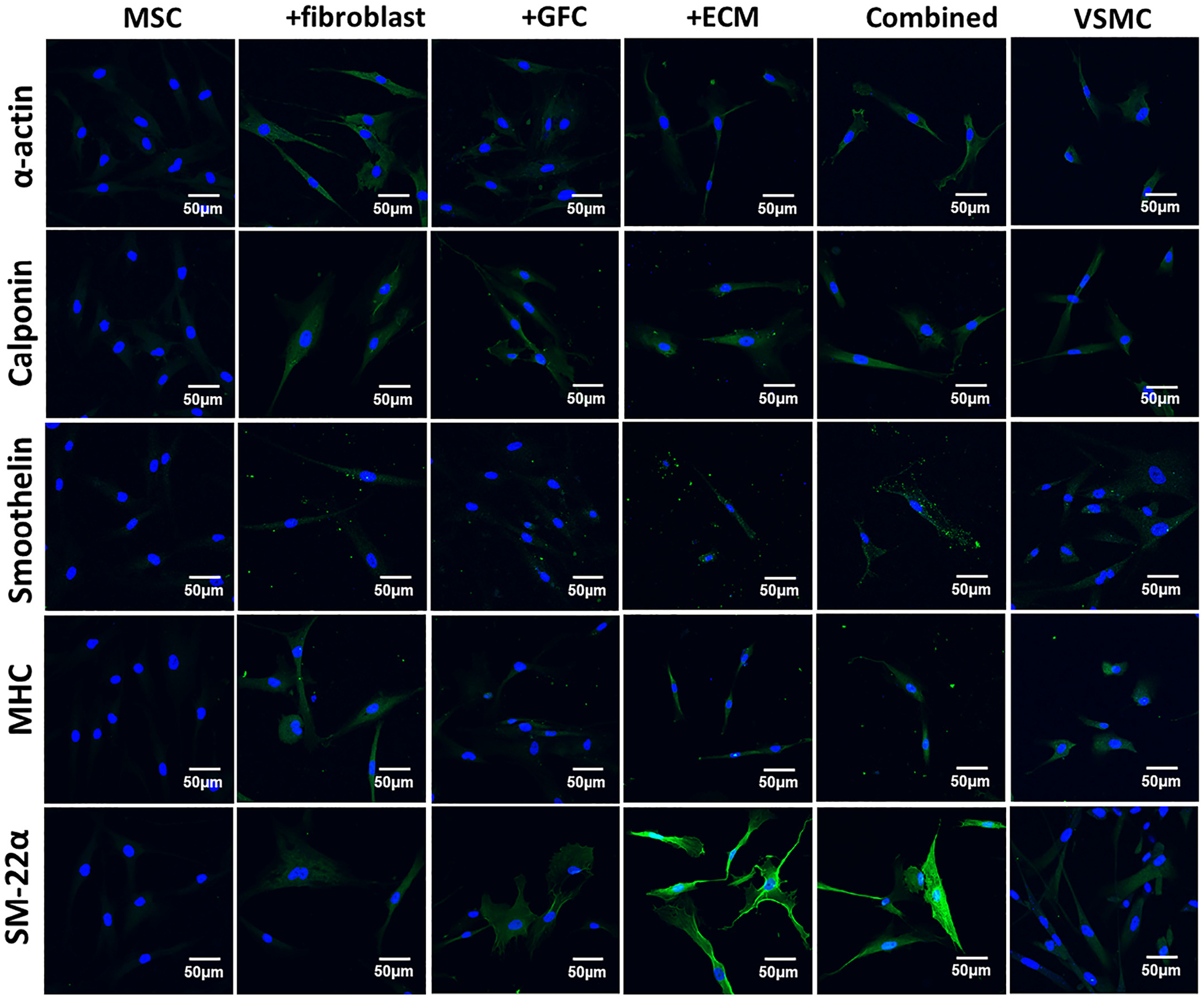
Fig. 4.

Immunofluorescence staining for VSMC-specific proteins including α-actin, calponin, smoothelin, myosin heavy chain (MHC), and smooth muscle protein 22α (SM-22α) in MSCs under different culture conditions for 14 days. Cell nuclei were counterstained with Hoechst 33342. Compared to MSCs, the fluorescence intensity (expression) of marker proteins in the differentiated cells induced by ECM scaffold, fibroblasts-coculture, GFC, or combined 3D coculture model were significantly increased. Fibroblasts and ECM appeared to be stronger regulators than GFC on the expression of α-actin, calponin, smoothelin, and MHC. In addition, the cells under ECM and combined 3D coculture condition demonstrated much higher fluorescence intensity for SM-22α than other experimental groups. The images represent at least three independent experiments.
