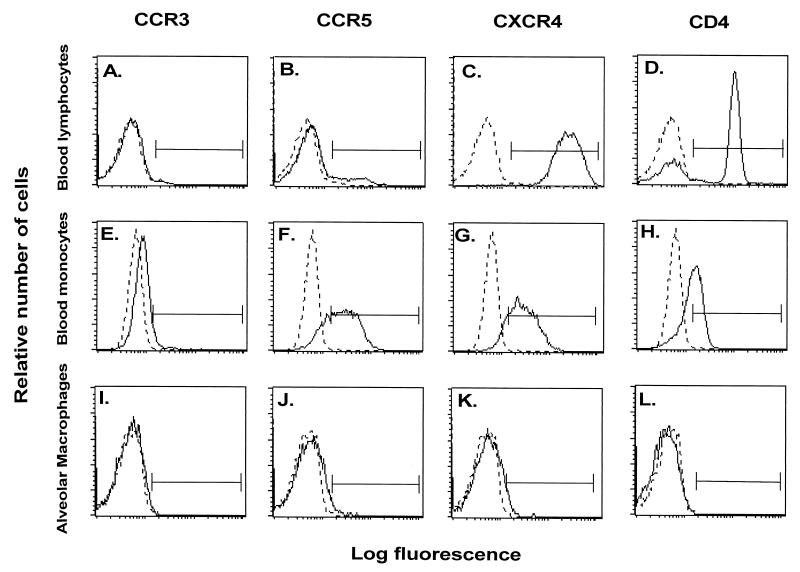
FIG. 2.
Flow cytometry evaluation of expression of chemokine receptors on AM. AM were obtained by bronchoalveolar lavage and stained with anti-CCR3, CCR5, or CXCR4 monoclonal antibodies (followed by FITC-labeled anti-IgG) or FITC-conjugated anti-CD4. Blood monocytes and lymphocytes obtained from the same donors were evaluated in parallel. Shown are representative samples from one individual of AM (panels A to D), PBM (panels E to H), and PBL (panels I to M). In addition to coreceptor staining, AM were double stained with PE-labeled HLA-DR, blood monocytes were double stained with PE-labeled CD14, and lymphocytes were double stained with PE-labeled CD3. The histograms shown represent the cells selected by these markers. IgG-irrelevant controls for each antibody are depicted by the dotted lines. Surface expression of CCR3 (panels A, E, and I); CCR5 (panels B, F, and J); CXCR4 (panels C, G, and K); and CD4 (panels D, H, and L) is shown. The solid horizontal line represents the region selected for quantification.

