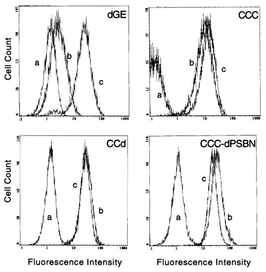
FIG. 4.
FACS analysis of cell surface markers on yolk sac cells transformed by dGE, CCC, CCd, and CCC-dPSBN. The same representative set of Myb-transformed yolk sac cells shown in Fig. 3 were stained with antibodies specific for cell surface markers. The antibodies 1C3 and HLO72 were used to detect granulocytic and monocytic lineages, respectively. Fluorescence profiles are shown after the cells were stained with wash medium control (a), 1C3 (b), or HLO72 (c).

