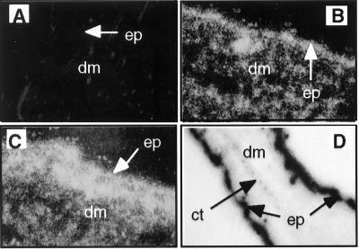
FIG. 1.
Examples of skin sections analyzed for E6 mRNA by in situ hybridization with a specific antisense RNA probe. (A to C) Dark-field images (magnification, ×400) of skin sections from nontransgenic (A), K14E6 transgenic line 5718 (B) and K14E6 transgenic line 5737 (C) mice. Bright areas indicate strong hybridization to an E6-specific probe. Note that signal is brightest in the epidermis (ep) but also present in underlying epithelial structures (hair follicles) in the dermis (dm). (D) Bright-field image (×100) of an ear section from K14E6 line 5737. Dark areas in the epidermis of both sides of the ear indicate strong hybridization to the E6-specific probe. Note absence of signal in the underlying dermis and cartilage (ct).

