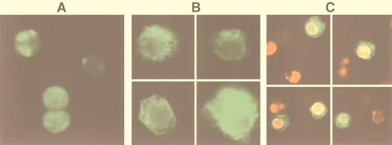
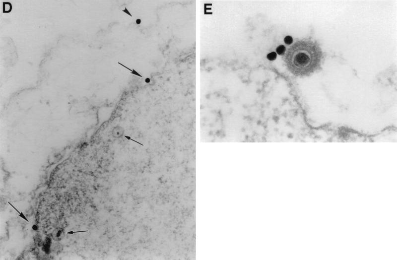
FIG. 2.
Visualization of PF-8 and gpK8.1 in BCBL-1 cells. Cells stimulated for 2 days with 30 nM TPA were fixed and permeabilized. (A and B) For IFA, cells were labeled with anti-PF-8 or anti-gpK8.1 MAbs followed by FITC-conjugated secondary Abs. The cells were centrifuged onto glass slides and photographed through a fluorescence microscope. Expression of PF-8 (A) and gpK8.1 (B) are shown. Magnification, ×245. (C) For colocalization studies, cells were labeled with both anti-PF-8 and anti-gpK8.1 MAbs followed by PE- and FITC-conjugated isotype-specific secondary Abs. Expression of PF-8 (red), gpK8.1 (green), colocalized PF-8 and gpK8.1 (yellow) are shown. Magnification, ×155. (D and E) For electron microscopy studies, cells were labeled with anti-gpK8.1 MAbs followed by gold-coupled secondary Abs. (D) Three gold beads are seen on a portion of a permeabilized cell, two attached to the nuclear membrane (arrows) and one on the plasma membrane (arrowhead). Note the moderately electron-dense granular material under the nuclear membrane and two nearby nucleoids (small arrows). Magnification, ×52,000. (E) Three 40-nm gold beads are associated with the surface of a typical mature HHV-8 virion. The nearby plasma membrane is free of label. Magnification, ×110,000. Data shown are representative.


