FIG. 3.
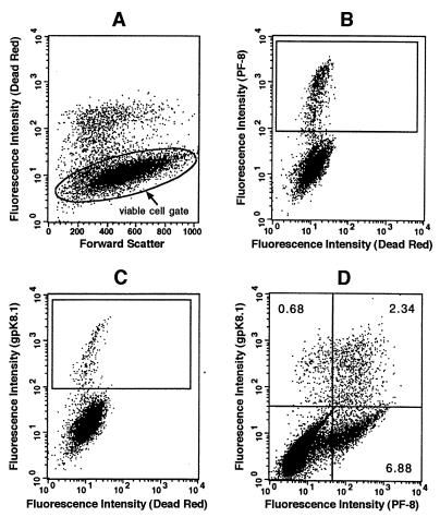
Detection of PF-8 and/or gpK8.1 in single cells by flow cytometry. (A to C) TPA-stimulated BCBL-1 cells were labeled with Dead Red, washed, fixed, and permeabilized. The cells were labeled with anti-PF-8 or anti-gpK8.1 MAb followed by FITC-conjugated secondary Abs. They were examined for fluorescence by flow cytometry. Dead Red-negative cells (representing the viable cell population) were gated (A) and examined for PF-8 (B) or gpK8.1 (C) expression. The numbers of cells in the defined regions were used for quantification and are shown in further experiments. No PF-8- or gpK8.1-expressing cells were observed when isotype control antibodies or HHV-8-negative cell lines were used (results not shown). (D) For coexpression experiments, cells were double labeled with anti-PF-8 and anti-gpK8.1 MAbs followed by PE- and FITC-conjugated isotype-specific secondary Abs. The numbers of viable cells expressing only PF-8, only gpK8.1, or both PF-8 and gpK8.1 are noted in the relevant quadrants. Data shown are representative of numerous experiments.

