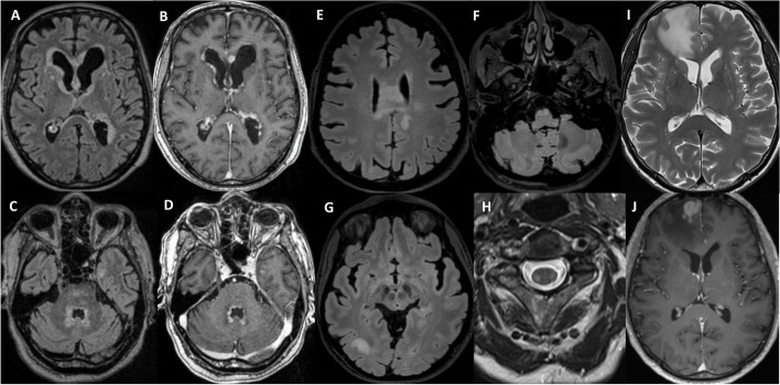
Fig. 1.
Brain MRI of the three cases. Case 1: Initial MRI with periventricular T2/FLAIR hyperintensities (A, C) and nodular gadolinium enhancement (B, D). Case 2: Brain MRI with T2/FLAIR hyperintensities of the corpus callosum extending to the corona radiata (E), of the right parieto-occipital subcortical white matter (G) and of the bilateral pyramids (F). Spinal MRI with bilateral lateral columns T2 hyperintensities (H). Case 3: Brain MRI with T2 hyperintense fronto-sagittal lesion and perilesional edema (I) and strong gadolinium enhancement (J)

