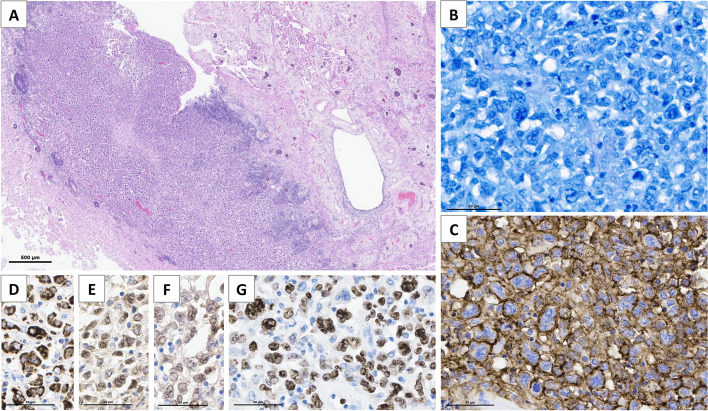
Fig. 2.
Brain autopsy findings in patient 1. Thickening of periventricular areas that are infiltrated by a diffuse population of large tumor cells without glandular or squamous differentiation (A, Hematoxylin–Eosin staining). The tumor is composed of a diffuse proliferation of large cells with a high nucleo-cytoplasmic ratio, mono- and multilobated nuclei, numerous mitoses (B, Giemsa staining) and high proliferation index Ki67/MIB1 near 80% (G). Tumor cells express CD20 (C), with coexpression of Bcl2 (D), Bcl6 (E) and MUM1 (F), and are monotypic for immunoglobulin light chain lambda, consistent with the diagnosis of primary diffuse large B-cell lymphoma of CNS (EBV negative, not shown)

