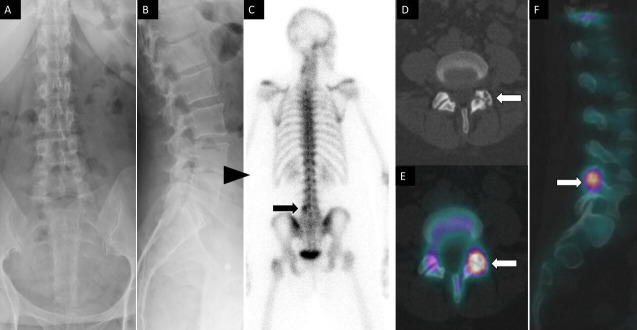
Figure 1.
A 52-year-old woman had lumbar back pain. Anteroposterior (A) and lateral (B) spine radiographs show mild multilevel degenerative disc narrowing most notable at L4 to L5 (arrowhead). Posterior planar 99m-methyl diphosphonate bone scan image (C) shows focal increased osteoblastic activity within the left lateral aspect of L5 vertebra. Axial noncontrast computed tomography (CT) (D) and fused axial (E) and coronal (F) single photon emission CT with CT images demonstrate increased osteoblastic activity within the left L4 to L5 facet joint with associated subchondral cysts and joint space narrowing compatible with degenerative facet arthropathy.

