
Figure 3.
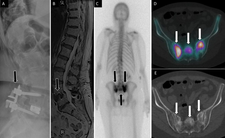
71-year-old woman with low back, buttock, and bilateral leg pain. Lateral (A) spine radiograph shows L4 to L5 instrumentation with prior interspinous spacer and unilateral pedicle screws with interbody device (arrow) without evidence of complications. Lumbar spine T2 sagittal magnetic resonance image (B) shows no central canal stenosis, also seen on axial (not displayed) and metal artifact at L4 to L5 (arrow) related to instrumentation. Posterior planar 99mTc-MDP bone scan (C) and axial fused single photon emission computed tomography with computed tomography (D) images show H-shaped radiotracer uptake within the sacrum, with associated sclerotic changes on computed tomography (E), compatible with sacral insufficiency fracture.