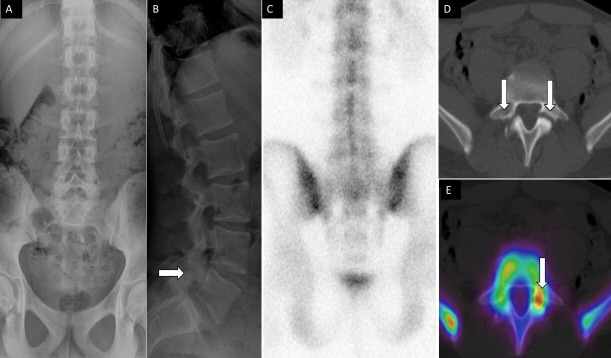
Figure 5.
A 14-year-old male football player presenting with low back pain. Anteroposterior (A) and lateral (B) spine radiographs show subtle lucency on the lateral view within the L5 pars interarticularis (arrow). Posterior planar 99mTc-MDP bone scan image (C) was unremarkable. Axial noncontrast computed tomography (CT (D) and fused single photon emission CT with CT (E) images demonstrate bilateral pars defects, with asymmetric increased activity within the left pars defect, respectively.

