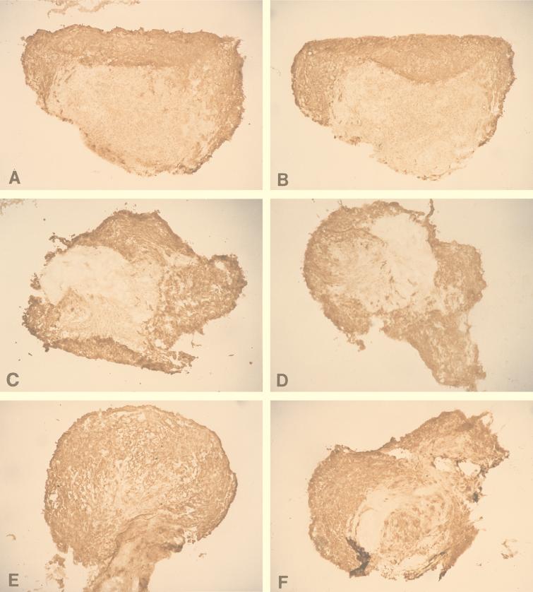
FIG. 6.
Sections through DRG (isolated from the thoracic region) from HSV-infected DRG-EC cultures at 36 (A and B), 48 (C and D), and 72 (E and F) hpi stained for HSV gC antigen. Human anti-gD (A, C, and E) or control medium (B, D, and F) was added to the outer chamber immediately after infection and left for 12 h. At 36, 48, and 72 hpi, snap-frozen (for 30 s in liquid nitrogen) HSV-infected and mock-infected DRG were mounted on a freezing cryotome (Shandon E-600) at −20°C and sectioned (perpendicularly to the coverslip) into 5-μm-thick sections. The sections were air dried on glass slides, stained with murine anti-gC1 antibody (1:100) (Goodwin Institute for Cancer Research) for 45 min, washed with HBSS and double-distilled water, and stained with biotinylated sheep anti-mouse antibody (Biosource International) (dilution, 1:200) for 45 min at RT. After two washes, sections were treated with streptavidin-horseradish peroxidase conjugate (Biosource International) at a 1:4,000 dilution. The proportion of all DRG neurons which were gC antigen positive was quantified in frozen sections of the whole mounted DRG (as described in Materials and Methods). Actual size of frozen DRG, 1.2 to 2.2 mm.

