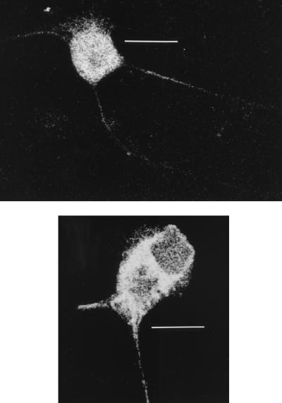
FIG. 7.
Confocal micrographs of HSV-infected neurons stained for gC antigen at 15 hpi and after addition of human anti-gD monoclonal antibody (top) or control medium (bottom). The HSV inoculum (5 TCID50/cell) was aspirated after 1 h of incubation, and the cells were carefully washed once with HBSS. The HSV-infected or mock-infected dissociated neuronal cultures, incubated with a 1:2,500 dilution (400 ng/ml) of human anti-gD antibody, were fixed in 2.5% formaldehyde (ProSci Tech) in Sorensons buffer (pH 7.4) for 30 min and permeabilized with 0.1% Triton X-100 (Sigma) in PBS for 20 min. Nonspecific staining was blocked by incubation with 5% mouse serum in HBSS for 15 min. The cells on coverslips were then incubated with fluorescein isothiocyanate-conjugated anti-gC1 antibody (Syva Microtrak) (1:100 dilution), rinsed three times with HBSS, and mounted in mounting fluid (Syva Microtrak). Stained neurons were examined with a Bio-Rad MRC 600 confocal microscope. Note the similar distributions of gC antigen in the axon and cytoplasm in both micrographs. Bars, 40 μm (top) and 20 μm (bottom).

