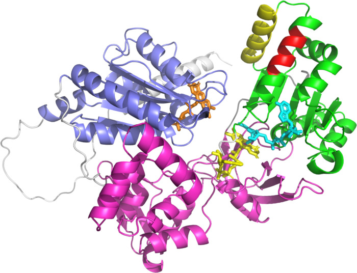
Fig 6. AlphaFold model of LdP450R1 in closed conformation.
The FMN, FAD and NADPH binding domains are highlighted in blue, magenta and green, respectively. The N-terminal membrane attachment domain is highlighted in grey. FMN (orange), FAD (yellow) and NADPH (cyan) are shown in stick representation with binding modes modelled from the rat P450R structure (1J9Z.pdb). Helix 21 (H21), known to directly interact with partner CYPs is highlighted in yellow. Amino acids 605–612, deleted in our AmB R1, form part of helix 20 (H20). Deleted amino acids are highlighted in red.

