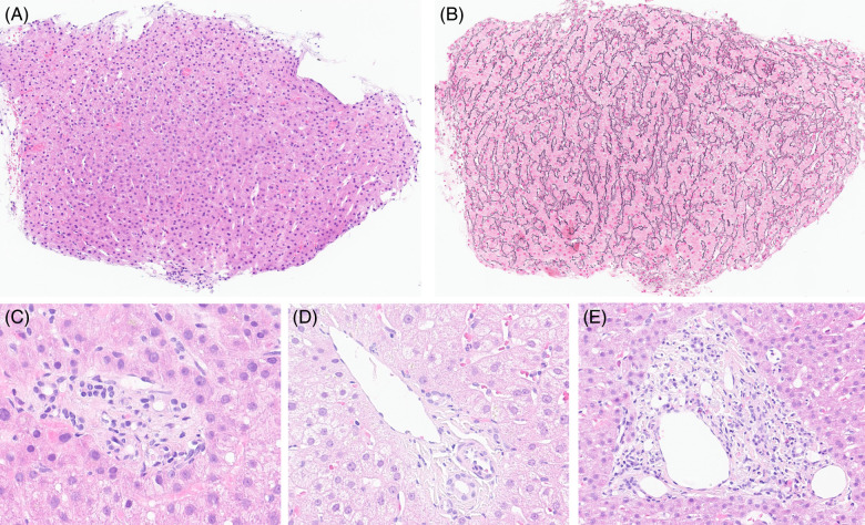
FIGURE 2.
Histological features of PSVD observed in our patient cohort. (A) and (B) Nodular regenerative hyperplasia, with parenchymal nodularity highlighted by reticulin stain in panel B (× 10). (C) Portal vein stenosis, characterized by a portal space with no clearly visible portal venous branch (× 40). (D) Herniated portal vein in direct contact with periportal parenchyma (× 40). (E) Hypervascularized portal space with multiple small-caliber venous branches (× 20). Abbreviation: PSVD, porto-sinusoidal vascular disorder.

