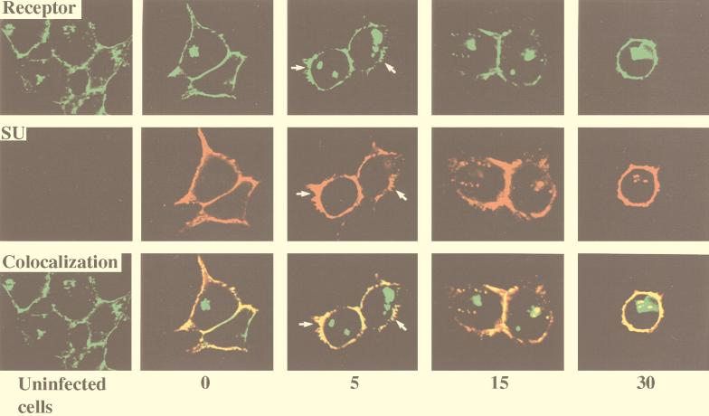
FIG. 3.
Colocalization of SU and MCAT-1-GFP in 293/MCAT-1-GFP cells after MoMuLV binding and following incubation at 37°C for different time periods. 293/MCAT-1-GFP cells were incubated with MoMuLV at 37°C for 0, 5, 15, or 30 min. Then cells were fixed, permeabilized, and stained with anti-SU (83A25), biotinylated goat anti-rat IgG secondary antibody, and Cy3-conjugated streptavidin. Color photomicrographs were produced with a Sony printer connected to the video output of the Zeiss confocal microscope. Arrows indicate significant membrane disturbance.

