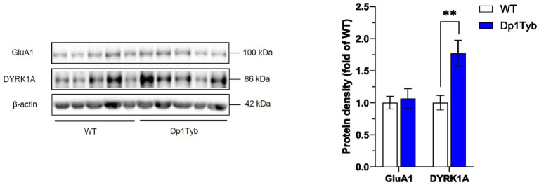
Figure 7.
Increased DYRK1A abundance in HPC cytosol from Dp1Tyb male mice. HPC cytosolic extracts from Dp1Tyb and WT mice were analyzed by immunoblotting for GluA1 and DYRK1A. Example immunoblot is shown on the left and mean ± SEM protein abundance on the right, normalized to β-actin and then to the mean signal in WT mice. Immunoblot shows analysis of HPC extracts from 5 mice of each genotype. Mutant Dp1Tyb mice displayed higher DYRK1A expression in hippocampal cytosol compared to WT littermates (Student’s t-test, **p < 0.01). 21-month-old Dp1Tyb: n = 6 WT, 5 Dp1Tyb.

