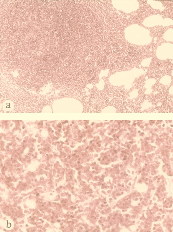
FIG. 3.
Histological examination of a representative tumor detected in a 7-week-old nu/nu rat after 3 weeks of subcutaneous inoculation of F344-S1 cells. Hematoxylin-eosin staining. (a) Low magnification of a tumor in the lung (×75). (b) High magnification of tumor cells in the lymph node. Note the polygonal tumor cells which contain ample cytoplasm and a large nucleus with a prominent nucleolus (×300).

