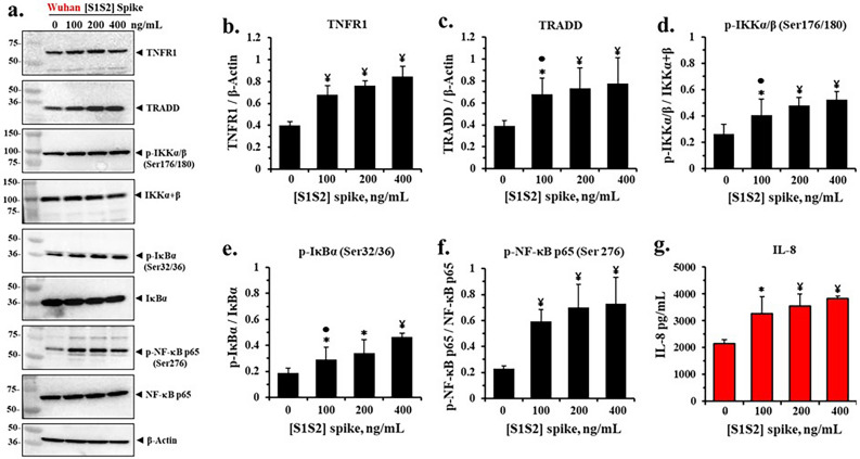
Figure 1.
SARS-CoV-2 [S1S2] spike protein activates pro-Inflammatory TNFα/NF-κB signaling proteins and IL-8 expression in differentiated BCi.NS1.1 (d-BCi) epithelia. The apical surfaces of the ALI differentiated dBCi epithelia were treated with different concentrations of Wuhan-Hu-1 [S1S2] spike protein for 4 h, washed and then incubated for an additional 20 h at the Air–Liquid-Interface (ALI) condition. (a) Representative western blotting images showing the expression levels of TNFR1, TRADD, phospho-IKKα/β (Ser 176/180), IKKα + β, phospho-IκBα (Ser 32/36), IκBα, phospho-NF-κB p65 (Ser 176), NF-κB p65, and β-Actin. β-actin was used for equal loading of protein. (b–f) Quantification by densitometric analysis for the data in part (a) using BioRad Image Lab software. The data are expressed as mean ± SD (N = 5). (g) IL-8 expression in the subphase of the ALI cultures. Data are presented as means ± SD (N = 3). Statistical p values were determined with a one-way ANOVA, followed by both Holm’s and Dunnett’s post-hoc tests comparing each mean to the medium control. For the Holm test, *, p < 0.05; and ¥, p < 0.01. Dunnett’s tests were consistently significant (p < 0.05), except as noted (•).

