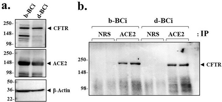
Figure 5.
ACE2 and CFTR co-immunoprecipitate in both basal cells and differentiated BCi.NS1.1 (d-BCi) epithelia. (a) A representative western blot image shows both untreated basal (b-BCi) cells and differentiated (d-BCi) cells expressing CFTR and ACE2. β-actin was used for equal loading of protein. (b) A representative western blot image showing CFTR co-immunoprecipitating with ACE2 from both basal (b-BCi) and differentiated (d-BCi) epithelia lysates. Normal rabbit serum (NRS) was used as the control. Western blot data represent the results of three independent experiments.

