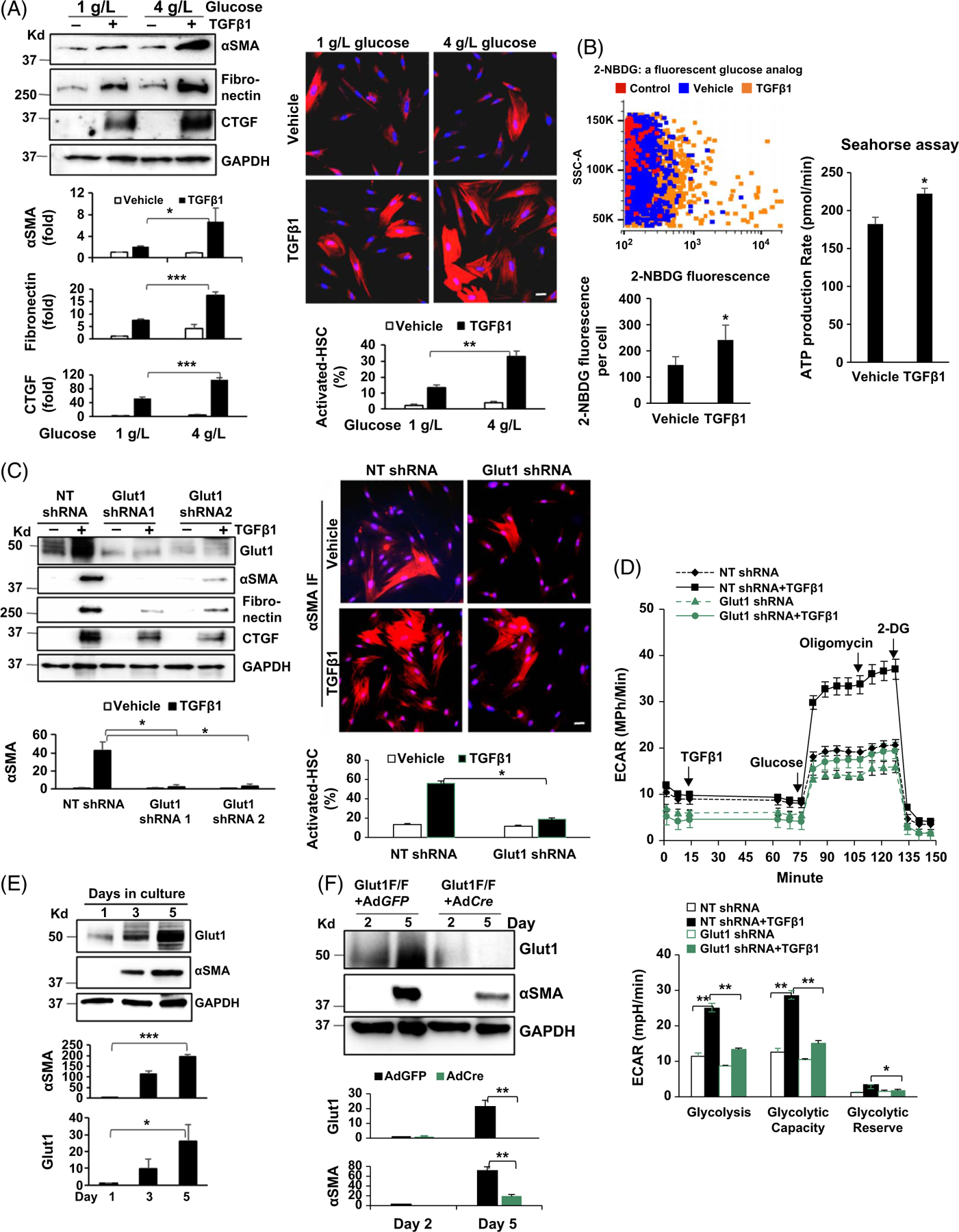
FIGURE 1.

Myofibroblastic activation of HSCs is promoted by Glut1. (A) Left, primary human HSCs in different glucose concentrations were stimulated with TGFβ1 (5 ng/mL) for 24 hours and collected for WB analysis. TGFβ1 upregulation of the stellate cell activation markers was potentiated by 4 g/L glucose compared to 1 g/L glucose. *p < 0.05 ***p < 0.001 by ANOVA, n = 3 repeats. Right, HSCs as described above were collected for αSMA immunofluorescence (IF). The rate of activated HSCs/myofibroblasts was increased by 4 g/L glucose. **p < 0.01 by ANOVA, n = 12 microscopic fields per group, each containing more than 100 cells. Bar, 20 μm. (B) Left, HSCs were incubated with 2-NBDG and 2-NBDG fluorescence within the cells was quantified by flow cytometry. TGFβ1 (5 ng/mL) promoted glucose uptake by HSCs. *p < 0.05 by t-test, n > 3,000 cells per group. Right, Agilent Seahorse XF96 ATP Rate Assay was performed. ATP production was increased by TGFβ1 stimulation (5 ng/mL). *p < 0.05 by t-test, n = 5. (C) HSCs expressing NT shRNA, Glut1 shRNA1, or Glut1 shRNA2 by lentiviral transduction were stimulated with TGFβ1 and collected for WB and αSMA IF. Glut1 knockdown suppressed myofibroblastic activation of HSCs induced by TGFβ1. *p < 0.05 by ANOVA, n = 3 repeats for WB; n = 12 microscopic fields per group for IF, each containing more than 100 cells. Bar, 20 μm. (D) Real-time ECAR was obtained by the Agilent Seahorse Glycolysis Stress test. The times when TGFβ1, glucose, oligomycin, and 2-DG were added are shown. TGFβ1 (5 ng/mL) promoted glycolysis in control HSCs, but not in Glut1 knockdown HSCs. *p < 0.05 **p < 0.01 by ANOVA, n = 5. (E) Murine HSCs were collected for WB. Glut1 expression by HSCs increased time-dependently similar to αSMA expression. *p < 0.05; ***p < 0.001 by ANOVA, n = 3 repeats. (F) HSCs isolated from Slc2A1/Glut1 floxed mutant mice were transduced with GFP adenoviruses (control) or Cre adenoviruses. Cre/LoxP-mediated Glut1 gene deletion suppressed murine HSC activation. **p < 0.01 by ANOVA, n = 3 repeats. Abbreviations: αSMA, alpha-smooth muscle actin; Cre/LoxP, Cre recombinase/LoxP sequence derived from bacteriophage P1; 2-DG, 2-deoxy-D-glucose; ECAR, extracellular acidification rate; Glut1, glucose transporter 1; IF, immunofluorescence; NBDG, 2-deoxy-2-[(7-nitro-2,1,3-benzoxadiazol-4-yl) amino]-D-glucose; NT shRNA, nontargeting short hairpin RNA; TGFβ1, transforming growth factor-beta 1; WB, western blot.
