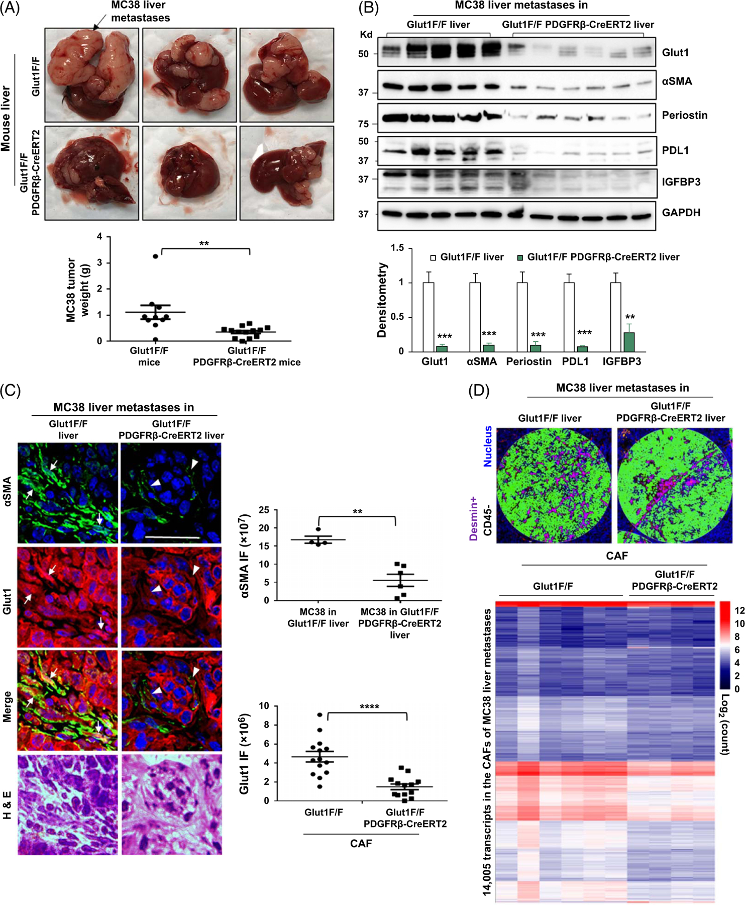
FIGURE 6.

HSC-specific Glut1 knockout by Cre/LoxP suppresses CAF activation, CAF transcriptome, and colorectal liver metastasis in mice. (A) MC38 murine colorectal cancer cells were implanted into littermate-matched Glut1F/F (control) and Glut1F/PDGFRβ-CreERT2 mice by portal vein injection. Glut1F/PDGFRβ-CreERT2 mice developed fewer MC38 liver metastases compared to control mice. **p < 0.01 by t-test, n = 10, 14. (B) WB with tumor lysates revealed that protein levels of Glut1, αSMA, periostin, PD-L1, and IGFBP3 were reduced in liver metastases of Glut1F/PDGFRβ-CreERT2 mice compared to those of control mice. **p < 0.01; ***p < 0.001 by ANOVA, n = 5, 6. (C) Double IF revealed that αSMA IF was reduced in MC38 liver metastases of Glut1F/PDGFRβ-CreERT2 mice compared to that in MC38 liver metastases of control mice (green). **p < 0.01 by t-test, n = 4, 6 tumors. Glut1 IF was also reduced in Glut1 F/PDGFRβ-CreERT2 CAFs (arrowheads) compared to Glut1F/F CAFs (arrows) (red). ****p < 0.0001 by t-test, n = 14, 13. Bar, 50 μm. (D) Upper, 2 example ROIs within MC38 liver metastases selected for transcriptomic profiling are shown. Anti-desmin IF was used to label the CAFs (purple), and anti-CD45 IF was used to label immune cells (orange). Desmin+CD45- areas were selected for analysis. Lower, the transcripts detected from the CAFs of different tumor sections are shown by a heatmap with a scale bar on the right. Abbreviations: αSMA, alpha-smooth muscle actin; CAF, cancer-associated fibroblast; Cre/LoxP, Cre recombinase/LoxP sequence derived from bacteriophage P1; Glut1, glucose transporter 1; IF, immunofluorescence; IGFBP3, insulin-like growth factor–binding protein 3; PDGFRβ, platelet-derived growth factor receptor beta; PD-L1, programmed death-ligand 1; ROI, regions of interest; WB, western blot.
