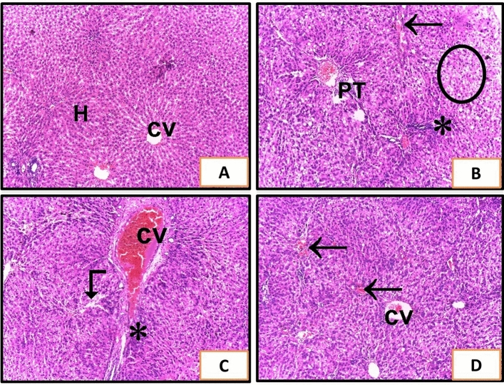
Figure 2.
Liver sections of rats from different experimental groups. (A) The control group (G1) and (G2) received sildenafil and showed a normal histological features of the liver with no abnormalities. Cords of hepatocytes (H) radiating from the central vein (CV). (B,C) show liver of G3 rats that received CCl4. (B) Shows sever degenerative changes with distorted histological architecture, vacuolization (circle), congested portal tract vasculature (PT), large inflammatory cells infiltrates (asterisk), and congested sinusoids (arrow). (C) Shows Sever congested central vein (cv), dilated sinusoids (angled arrow), and large inflammatory cells infiltrates (asterisk). (D) G4 received sildenafil and CCl4 and showed improvement of hepatic architecture with congestions between some hepatocytes (H&E ×100).

