FIG. 1.
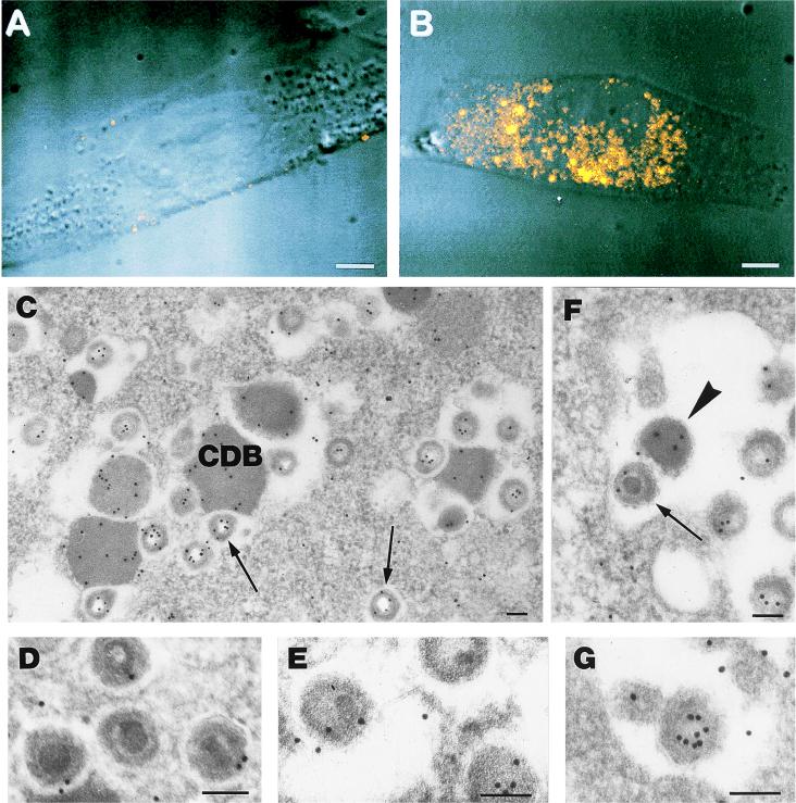
(A and B) Confocal microscopy of HEL cells labeled with anti-pUL25 antibody. (A) Mock-infected cells show a very faint background signal. (B) At 96 h after infection with CMV, the fluorescence due to the anti-pUL25 antibody in the cytoplasm indicates the presence of the protein in cytoplasmic bodies of different sizes. Bar = 5 μm. (C through G) Electron microscopy of HEL cells stained with anti-pUL25 antibody 120 h after infection. (C) Gold particles are present on CDB and on virion particles generally devoid of a core. The majority of the gold particles are localized along the tegument (arrows). (D and E) High magnification of intracytoplasmic virions in which the core is visible. The labeling is less intense on the tegument than in the virions from which the core has been removed by the section etching, and the tegument is more available to antibody binding (panel C). (F) Extracellular enveloped virions are labeled on the tegument (arrow). An immunolabeled dense body is also present in the extracellular space (arrowhead). (G) Detail of an extracellular virion which is tangentially sectioned, thus not allowing the detection of the core, which presents intense labeling on the tegument. Bar = 0.1 μm.

