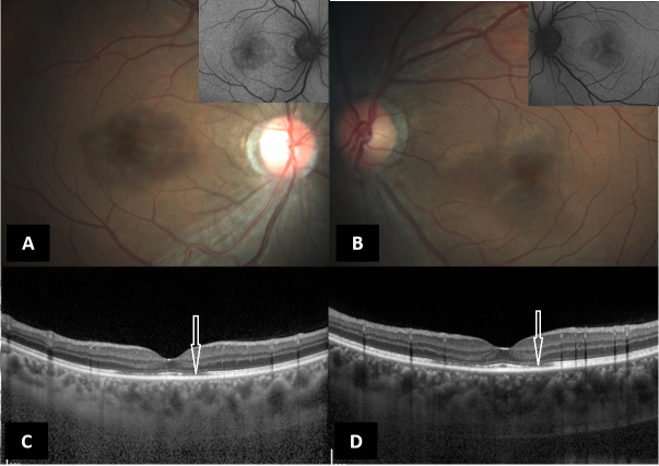
Figure 1.
(A & B) Color fundus photograph (Zeiss, Carl Zeiss Meditech, Germany) of the right and left eyes, respectively, showing ass RPE alterations with corresponding hypo autofluorescence (inset). (C & D)Spectral domain optical coherence tomography (Spectralis, Heidelberg Engineering, Germany) of the right and left eyes, respectively, showing parafoveal focal loss and thinning of the ellipsoid zone (white arrows).

