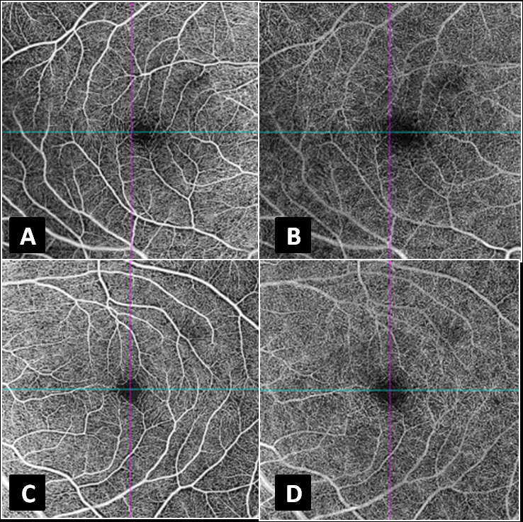
Figure 2.

(A & C) Swept source optical coherence tomography–angiography (DRI-Atlantis, Topcon, Oakland, NJ) of the right and left eyes, respectively, segmented at the superficial capillary plexus, showing no evident microcirculatory abnormality. (B & D) Swept source optical coherence tomography–angiography (DRI-Atlantis, Topcon, Oakland, NJ) of the right and left eyes, respectively, segmented at the deep capillary plexus, showing no evident microcirculatory abnormality.
