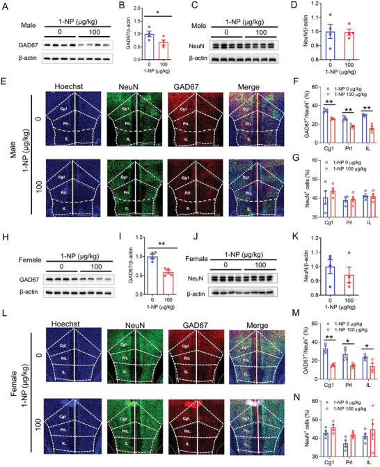
Figure 4.

Influence of maternal 1‐NP exposure on GAD67+ interneurons in weaning offspring. 10 pregnant mice orally received different dose of 1‐NP (0, 100 µg kg−1) daily from GD0 to GD17. All pregnant mice gave birth naturally. A–G) Male offspring were euthanized on PND28 and their mPFC was harvested. A,B) GAD67 was performed by Western blot. C,D) NeuN was examined through Western blot. E) GAD67+ and NeuN+ neurons in each subfield of the mPFC were analyzed using IF. F) The proportion of GAD67+ to NeuN+ neurons in each subfield of the mPFC. G) The percentage of NeuN+ neurons in each subfield of the mPFC. Original magnification: 400×. N = 4. *P < 0.05. **P < 0.01. H–N) Female offspring was sacrificed on PND28 and the mPFC was collected. H,I) GAD67 was measured by Western blotting. J,K) NeuN was analyzed using Western blotting. L) GAD67+ and NeuN+ neurons in each subfield of the mPFC were analyzed using IF. M) The proportion of GAD67+ to NeuN+ neurons in each subfield of the mPFC. N) The percentage of NeuN+ neurons in each subfield of the mPFC. Original magnification: 400×. N = 4. *P < 0.05. **P < 0.01. Cg1, cingulate cortex, area 1. PrL, prelimbic cortex. IL, infralimbic cortex.
