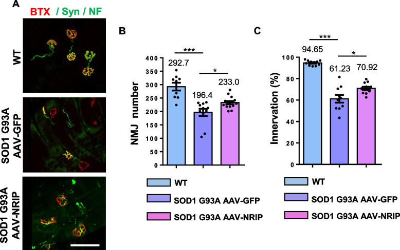
Fig. 5.
AAV-NRIP gene therapy increases NMJ number and axon terminal innervation at NMJ in SOD1 G93A mice. A Immunofluorescence analysis of NMJ and axon terminal innervation at NMJ. GAS muscles were incubated with α-BTX (red) and anti-Syn/NF antibodies (green). Scale bar: 100 μm. B Quantification of NMJ number in GAS muscles from WT mice (N = 9) and SOD1 G93A mice treated with AAV-GFP (N = 11) and AAV-NRIP (N = 14) at the age of 120 days. Quantification analysis of NMJ number was counted from the sum of three Sects. (30 μm thickness) of GAS muscles from each mouse. Data are presented as mean ± SEM. ***P = 0.0002 (WT vs. SOD1 G93A + AAV-GFP); *P = 0.0481 (SOD1 G93A + AAV-GFP vs. SOD1 G93A + AAV-NRIP); one-way ANOVA with Tukey’s post hoc test. C Axon terminal innervation at NMJ. The co-staining of α-BTX and synaptophysin indicated axon terminal innervation at NMJ of GAS muscles. WT mice (N = 11), SOD1 G93A mice infected with AAV-GFP (N = 10), and AAV-NRIP (N = 12) were analyzed. Quantification analysis of NMJ innervation is defined as the percentage of innervated NMJ to total NMJ number. Data are presented as mean ± SEM. ***P = 0.0002 (WT vs. SOD1 G93A + AAV-GFP); *P = 0.0183 (SOD1 G93A + AAV-GFP vs. SOD1 G93A + AAV-NRIP); one-way ANOVA with Tukey’s post hoc test

