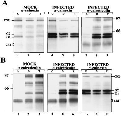
FIG. 1.
Coprecipitation of UUK virus spike proteins G1 and G2 with calnexin and calreticulin after solubilization with different detergents. Virus- or mock-infected BHK21 cells were labeled at 14 h postinfection for 10 min with [35S]methionine and then chased for 10 min. The cells were lysed in buffers containing different detergents (2% CHAPS [C], 1% digitonin [D], 1% Triton X-100 [T]) and processed for sequential immunoprecipitation. Lanes 1 to 3, proteins coprecipitated from uninfected cells; lanes 4 to 6, coprecipitation with calnexin (A) or calreticulin (B); lanes 7 to 9, proteins reprecipitated with anticalreticulin (A) or anticalnexin (B), respectively, from the supernatants remaining after three rounds of precipitations with the antisera used for lanes 4 to 6. Proteins were analyzed on an SDS–10% polyacrylamide gel under reducing conditions, followed by fluorography. The positions of calnexin (CNX), calreticulin (CRT; migrates as a double band), G1, and G2, as well as molecular weight markers (in thousands), are indicated.

